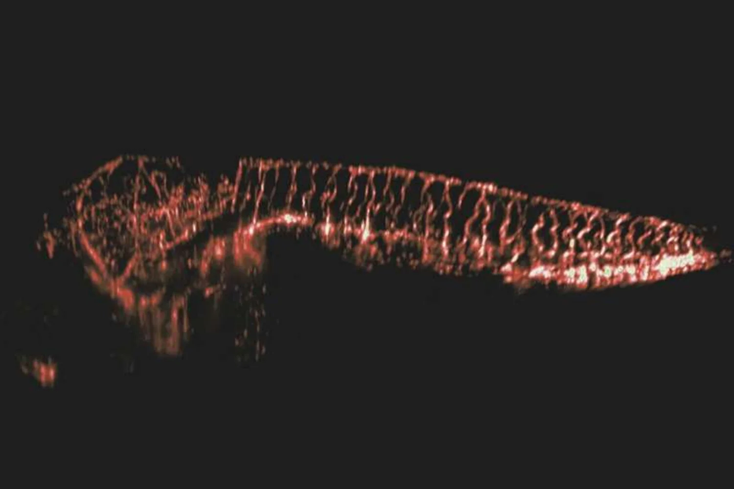
The scientists obtained a 3D model of living zebrafish larvae with the resolution of individual cells. An article about it was published in the journal Optica.
Danio rerio is a popular aquarium fish often used in biological experiments. Ji Yi of Johns Hopkins University and colleagues used its larvae to test the dynamic fluorescent microscopy system; at this time, the researcher observed the glow of an object from a laser aimed at it (living objects were injected with special fluorescent proteins). This method allows you to observe individual cells of living organisms, but like other microscopy methods, it has a limited field of view and does not allow you to see millimeter organisms as a whole.
The authors managed to circumvent this limitation with the help of a special optical element, the diffraction transmission grating. As a result, the scientists managed to obtain a three-dimensional image of 5.4 × 3.3 millimeters with a resolution of 2.5 × 3 × 6 microns. Thanks to this, they made volume recordings of the neural activity of the entire body of zebrafish larvae with a frequency of 2 Hz, which makes it possible to study the functioning of the nerve chains of vertebrates. The authors also demonstrated volume recordings of whole body blood flow at a frequency of 5 Hz, allowing to monitor the movement of cells within the entire circulatory system.
Source: Port Altele
