We associate the diagnosis of a disease with the moment when the doctor, sitting in front of us, tells us ‘you have a tumor’, ‘you don’t have a tumor’, ‘you have a clogged artery’, ‘you have a healthy heart’, ‘in a pneumonia”, “there is nothing in the lungs”.
An extraordinary number of these words, which sometimes make our heart beat faster, sometimes allow us to breathe easier, originate from data from imaging tests: X-rays, CT scans, ultrasounds, angiograms. , magnetic resonance imaging (MRI) ). The doctor tells us what we have or do not have based on what was possible to capture with a machine created by engineers, programmers, physicists and mathematicians. This means that, before becoming a clinical issue, medical diagnosis is a technological issue. Ultimately, the question is: what can the diagnostic equipment in question ‘see’?

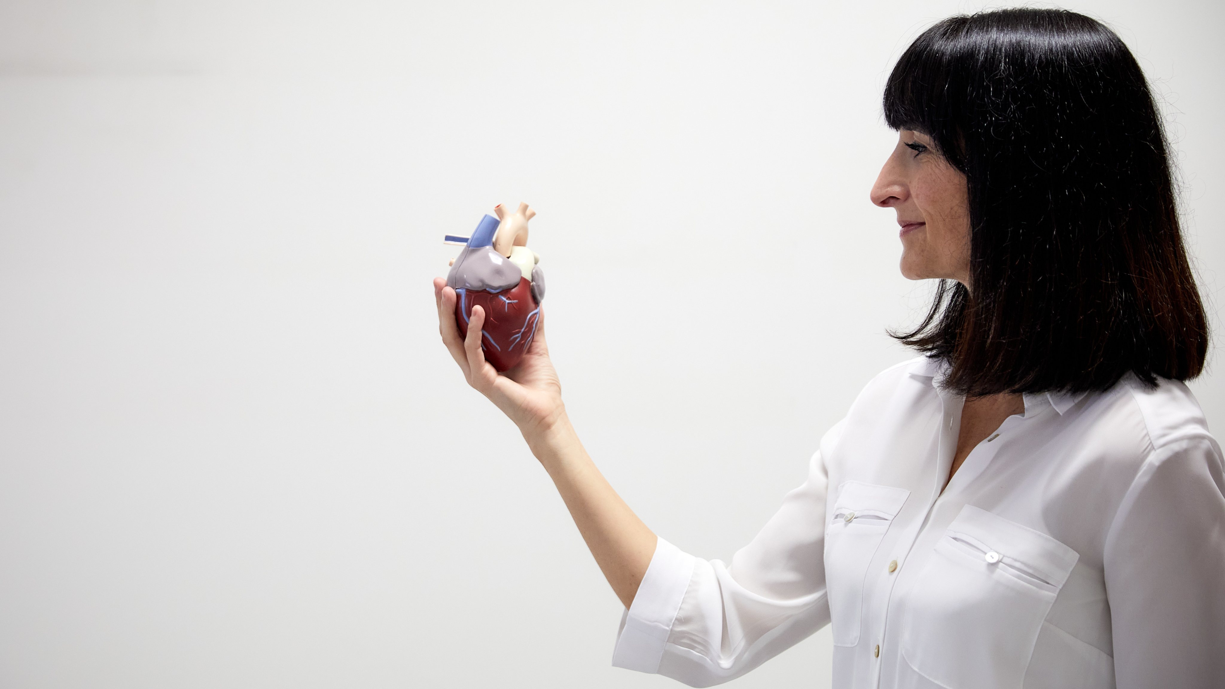
This is the question that researcher Teresa Correia, 40, from the Centro de Ciências do Mar do Algarve (CCMAR), has been working on for 15 years: developing ways that allow machines to see more and better. She has used her training in physics, mathematics, engineering and programming to improve the techniques of acquisition, reconstruction and correction of movement in medical imaging, specifically in Magnetic Resonance Imaging (MRI) applied to cardiovascular diseases. With her new project, financed by Fundação la Caixa, she aims to improve the diagnosis of coronary heart disease (CHD), the leading cause of death worldwide, which occurs when blood flow to the heart is limited.
“Coronary artery disease results from the accumulation of fatty plaques in the arteries that supply the heart, which makes it difficult for blood to pass through, causing the heart muscle, the myocardium, to receive less nutrients and oxygen,” explains the researcher. .
In the case of a total obstruction of the arteries, a myocardial infarction -or, as we usually call it, a heart attack- can occur, so it is important to have an early diagnosis and treatment of the disease to avoid serious or fatal complications. ”
Currently, the method used to detect the disease is coronary angiography, but the test has some limitations. “It’s expensive, invasive, it uses X-ray radiation and only detects arterial blockages, the disease is not visible at an earlier stage, which can occur at the level of the smallest blood vessels, the branches of these coronary arteries,” he explains. the investigator
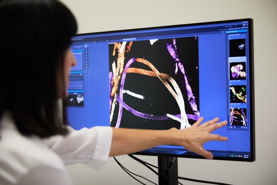
The most complete alternative is Cardiac Magnetic Resonance, a technique used –and recommended– to observe cardiac function, tissue viability and also the flow of blood through the heart, through the contrast medium administered. “But this pass takes less than a minute, which means we have to get a lot of information in a short time. That means we have to sacrifice something. Currently, we sacrifice image quality, which is relatively low by MRI standards, and we also cannot get a full image of the heart, just sections.”
In addition to the technical challenges, there is a problem for the patient that Teresa Correia knows well: it is necessary to hold your breath several times, so that the image does not come out ‘shaky’. “I’ve been inside an MRI machine several times, as a guinea pig, trying my own methods, and I know it’s hard to hold your breath. And I am a healthy person, for someone who is sick it will be more complicated. We often end up with images that are impaired by respiratory motion and may not be useful.”
The apnea required of the patient and the incomplete image with little detail are pertinent technical difficulties. But today, the biggest problem is the difficulties of human resources. “It is a complex technique and the interpretation of the data requires highly qualified personnel, with a long period of training. That is why the exam is only done in specialized centers, there are many places that have the technical capacity to do it, but do not have the human resources”.
There are many problems and Teresa Correia will try to solve them all: she proposes to change the way the machine acquires the data and the way it is presented. “That is why we have a collaboration with Philips Healthcare [uma das principais empresas de desenvolvimento de equipamentos de RM] this allows us to access the MR machine software and make changes”, explains the researcher. “We are going to combine data acquisition techniques with mathematical modeling of cardiac blood flow, image reconstruction, and also motion correction.”
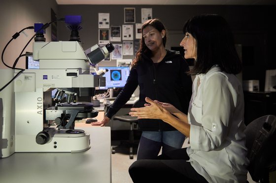
The goal is to obtain an image of the whole heart, and not just sections, with better quality and without the patient having to hold their breath. “If everything goes well, we will ensure that the machine obtains more and better information, reduces patient discomfort”, says Teresa Correia, but also facilitates the work of the person performing the exam, “be it because the machine does many of the it works automatically, requiring less training, either because it generates quantitative maps that facilitate the interpretation of the results.”
In addition to the collaboration with Philips Healthcare, the project will work in consortium with three other institutions. In Spain they have a research group from the Centro Nacional de Investigação Cardiovascular Carlos III, in Madrid, for clinical trials, and another from the University of Valladolid, which will provide experience in the development of movement correction techniques. In Portugal, it has the collaboration of the Associação do Instituto Superior Técnico de Investigación y Desarrollo to work on data acquisition.
Born in Olhão, Teresa Correia moved to Lisbon at the age of 17 to study at the Instituto Superior Técnico. Her inclination for science had been evident for some time and she even thought of studying Aerospace Engineering. The universe fascinated her-she still fascinates her-and she imagined working at the European Space Agency (ESA), but then she read an article in a newspaper about two former students of the Instituto Superior Técnico (IST) who were doing research in California . The article said that they had graduated in Physics and Technological Engineering. She had never heard of it before, but it sounded good and seemed more complete. Applied and entered.
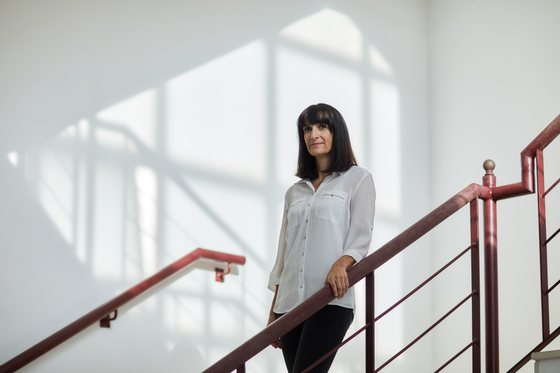
Happily I won the five hours by bus between Olhão and Lisbon. “I was fed up with a small environment and wanted to go to a big city,” she says. And Lisbon was big, but not big enough. Therefore, at the end of the course, she left for Copenhagen, where she stayed for about a year and a half. She did an Erasmus at the Niels Bohr Institute, at the University of Copenhagen and was also at Risø, the Danish national laboratory, where she had her first contact with research in the field of biomedicine. “However, in the end, Copenhagen also seemed like a very small city to me.” She then went to London to do a PhD in Medical Physics and Biomedical Engineering at University College London, where she worked for some years observing infant brain development through optical tomography. Subsequently, she moved to King’s College London, where she stayed until two years ago, researching MRI techniques applied to cardiac problems.
Until she began to rethink her priorities. “And after 15 years of living in a very big city, I realized that I wanted to live in a small town,” she confesses with a laugh. He wanted to return to the Algarve and wanted to create a center in the area of digital technologies, bioimaging and magnetic resonance that would contribute to the development of the region. She began to think about the importance of decentralizing and to question some preconceived ideas: “Why does this work have to be done in a big city and can’t it be done in a small town?”
“Before coming to the Algarve I thought: what do I need to make this work? And the answer was: a computer. I can do a lot of research with a single computer and the rest remotely through collaborations and travel when necessary.” It is precisely what you are doing. The Center for Marine Sciences of the Algarve (CCMAR) welcomed him. He uses his experience in the area of optical imaging to apply this technique to the study of marine biology and, at the same time, launches his dream of creating a reference center in the area of medical imaging near his homeland, through of funding from Fundação the Box. “One of the things that made me the happiest lately? See the publication of the results of the CaixaResearch de Investigação em Saúde 2022 contest marked on a map. Of the 13 projects in Portugal, most of the points are in Lisbon, Porto, Coimbra. But there is also one in the Algarve”. The dream of placing the region on the world map of diagnosis through bioimaging has already begun.
This article is part of a series on cutting-edge scientific research and is the result of a collaboration between the Observer, the “la Caixa” Banking Foundation and BPI. The project Direct quantitative assessment of heart disease with magnetic resonance imaging, led by Teresa Matias Correia, from CCMAR, was one of the 33 selected (13 in Portugal) -among 546 applications- to receive funding from the Barcelona-based foundation, within the framework of the 2022 edition of the Research Contest in Health CaixaResearch. The researcher received one million euros to develop the project for three years. Applications for the 2022 edition closed on November 15. The deadlines for the 2023 edition should be known in the first half of next year.
Source: Observadora
