Death is a deeply disruptive event for a living brain, to say the least. The cascade of effects that occurs when oxygen is lost sweeps like a tide over how our cells copy and translate our DNA to keep the lights on. Comparing postmortem brain tissue with samples from living patients has revealed, for the first time, significant differences in the ways RNA strands are altered, revealing new potential targets for disease diagnosis and treatment.
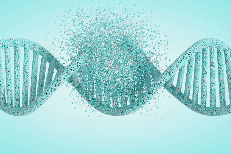
Researchers at the Icahn School of Medicine at Mount Sinai in New York focused on how specific adenosine (A) base codes in messenger RNA are replaced with a completely different base, inosine (I).
“Until now, studies of A-Z editing and its biological significance in the mammalian brain have been limited to post-mortem tissue analysis,” says genome expert Michael Breen. “By using fresh samples from living humans, we were able to detect important differences in RNA editing activity that previous studies based solely on post-mortem samples may have missed.”
To translate genes encoded by double-stranded DNA strands into functional proteins, biology must transcribe their sequences into a slightly different format than RNA. These “messengers” can then be translated into proteins using other RNA structures that link the building blocks of amino acids together.
Billions of years of evolution have taken advantage of this intermediate transcription and translation service to add an almost entirely new library of proteins. Like a rogue editor who changes the text of texts to serve entirely new purposes, cells can adjust a gene’s messenger RNA to meet entirely different needs.
Some species, particularly cephalopods, take RNA editing to a whole new level, rewriting their brains’ own genetic instructions to suit their circumstances.
In vertebrates like us, removal of the amino group, or “deamination,” of adenosine converts it to inosine, a base similar to guanine (G), resulting in an end product that is very different from what is usually encoded in a DNA gene library.
This modification of AI bases is carried out by adenosine deaminase, which acts on the RNA (ADAR) family of enzymes that play an important role in the formation of many different tissues, including the brain.
In fact, this process is so critical that errors in the editing process can lead to a variety of neurological disorders. To determine exactly how changes in specific copied genes translate into life-threatening conditions, researchers analyzed samples collected after death. As easy as these samples are to collect, they have a serious drawback.
“We hypothesized that molecular responses to postmortem hypoxic and immune responses could significantly alter the editing landscape from A to Z,” says lead study author Miguel Rodríguez de los Santos, a molecular biologist at Mount Sinai. “If we only examine postmortem tissue, this could lead to misunderstandings about RNA editing in the brain.”
In fact, brain tissue samples obtained from living patients at the time of surgical implantation of deep brain stimulation electrodes revealed significant differences in the activity of the two types of ADAR enzymes and in the regions where they were affected. The team’s analysis identified more than 72,000 regions in RNA strands where A-to-I editing occurred more frequently in samples from recently deceased patients compared with those collected from living patients.
But there were hundreds of sites where the opposite happened, where the editing process was more efficient in living brain samples. Although some regions have known brain plasticity functions, many need further research to understand the mechanisms involved.
“It is important to note that our findings do not negate, but rather provide, a missing context for the use of postmortem brain tissue in A-to-Z editing studies,” said Alexander Charny, one of the senior authors and a physician scientist at Mount Sinai.
“Understanding these differences helps improve our knowledge of brain function and disease through the lens of RNA editing modifications, potentially leading to better diagnostic and therapeutic approaches.” This study was published in: Nature Communication.
Source: Port Altele