
In a wild biological manipulation that seems straight out of science fiction, researchers have made parts of living mice transparent. Stanford University materials scientist Zihao Wu and colleagues have developed a biosafe dye that makes tissues transparent by affecting the ability of the fluids surrounding cells to scatter light.
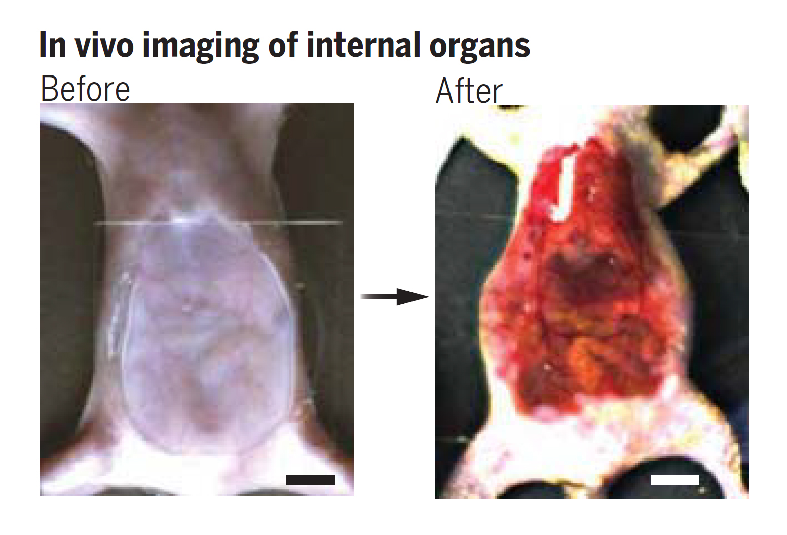
It is hoped that such strategies will allow researchers to clearly observe the functioning of the body’s internal organs as they function within a living body.
“In the future, this technology could make blood vessels more visible for blood sampling, make it easier to laser tattoo removal, or aid in the early diagnosis and treatment of cancer,” says Stanford University materials scientist Guosong Hong.
When light of a particular wavelength passes through materials with different refractive properties, it scatters in all directions, making the material appear opaque. For this reason in large part, even the thin layers of tissue that make up animal skin and the fluids that surround them are generally not transparent.
If such biological materials have the same refractive index, light rays can reflect from deeper tissues and pass smoothly over the boundary, providing a level of resolution that would otherwise be lost due to scattering. This already occurs naturally in some animals, including glass frogs (Hyalinobatrachium fleischmanni) and zebrafish (Danio rerio).
One way to do this in opaque tissues is to introduce a substance that absorbs incident light at a very specific wavelength. The mathematical relationship between the absorption and refraction of a material, based on the Kramers-Kronig relation, means that fine-tuning one property allows the other to be changed to a precise degree.
The researchers discovered that a food-safe dye called tartrazine can absorb some of the light of the desired color, allowing the researchers to change the refractive index of the fluid surrounding the cells and greatly reduce scattering.
“The dye is biocompatible; it is safe for living organisms,” Ou explains. “It is also very cheap and effective; we don’t need a lot of it to work.”
Applying a mixture of orange-yellow tartrazine dye and water to a mouse’s skin allowed engineers to see details of blood vessels and organs, and even observe muscle contractions in the animal’s digestive tract.
“It takes a few minutes for the transparency to appear,” Ou says. “The duration depends on how quickly the molecules diffuse into the skin.”
The dye can then be washed off, leaving the skin matte again. Any tartrazine that has penetrated deeper into the body will eventually be eliminated from the body.
“As an optician myself, I am amazed at how much they have achieved from the use of Kramers-Kronig junctions; it is a wonderful example of how fundamental optics knowledge can be used to create new technologies, particularly in biomedicine,” said Adam Wax, program manager at the US National Science Foundation, which supported the work.
But since human skin is about 10 times thicker than mouse skin, it’s not yet clear whether a similar method would work for us. The researchers are keen to investigate this further. The study was published on: Science.
Source: Port Altele